Abstract
Objective. To evaluate the effectiveness of balneotherapy (mud packs and sulfur baths) on patients with psoriasis and psoriatic arthritis (PsA).
Methods. One hundred and sixty-six patients with psoriasis and PsA were treated at the Dead Sea for a period of 3 weeks. The patients were divided into 2 groups. Both groups had a regular regimen of bathing in Dead Sea water and exposure to the sun’s ultraviolet rays. The study group, which consisted of 146 patients also was treated with mud packs and sulfur baths. The control group, which had no additional
therapy, consisted of 20 patients. The main clinical variables assessed were duration of morning stiffness, grip strength, activities of daily living, subjective patient assessment of disease severity, number of active joints, and number of effluent joints. Ritchie index, psoriasis area and severity index score, cervical, thoracic, and lumbar spine pain and limitations of movement.
Results. Statistically significant improvement was found in most variables in both groups. However, better results were observed in the study group. In 2 variables, reduction of spinal pain and range of movement in the lumbar spine, significant improvement (p < 0.001 and p = 0.022, respectively) was observed in the study group only.
Conclusion. Treatment of psoriasis and PsA at the Dead Sea area is very efficacious and the addition of balneotherapy can have additional beneficial effects on patients with PsA. Other controlled studies with longer follow-up periods are needed to verify our results. (J Rheumatol 1994:21:1305-9)
Key Indexing Terms:
PSORIASIS PSORIATIC ARTHRITIS DEAD SEA BALNEOTHERAPY
From the Rheumatology Unit and the Departments of Medicine D. Epidemiology and Dermatology, Soroka Medical Center of Kupat-Holim, Faculty of Health Sciences, Ben-Gurion University of the Negev, Beer- Sheva, and Hamei-Zohar Spa Clinic at the Dead Sea, Israel. S. Sukenik, MD. Professor, Head, Department of Medicine D; H. Giryes, MD, Resident in Dermatology; S. Halevy, MD, Associate Professor, Head, Department of Dermatology; L. Neumann, PhD, Associate Professor, Head, Department of
Epidemiology; D. Flusser, MD, Lecturer; D. Buskila, MD, Senior Lecturer, Head, Rheumatology Unit.
Address reprint requests to Dr. S. Sukenik, Department of Medicine D. Soroka Medical Center, Beer-Sheva, Israel 84101. Submitted July 6, 1993 revision accepted December 9, 1993
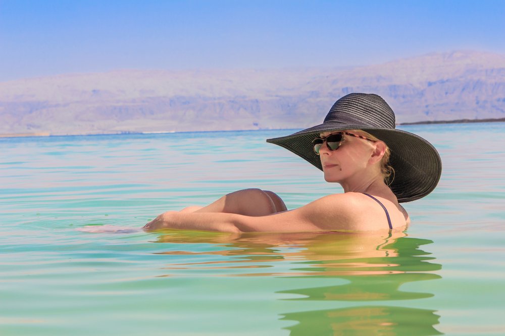
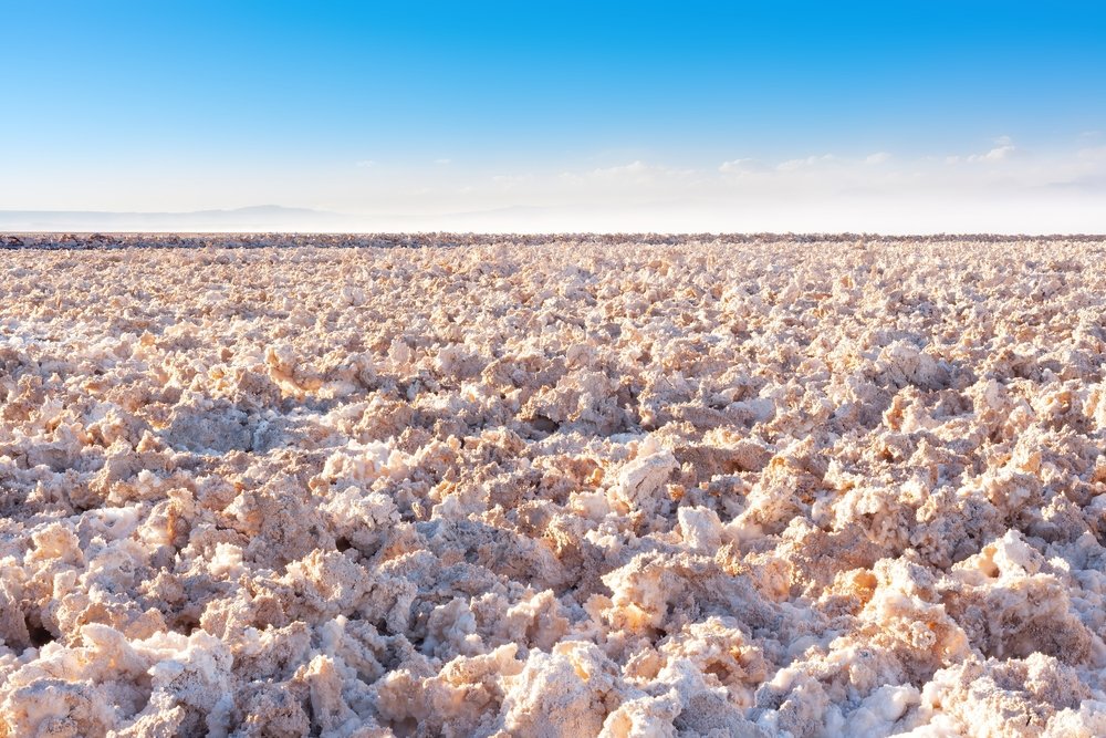
The role of balneotherapy and climatotherapy in rheumatology is a subject of much debate. In Israel, the main health resort area proving balneotherapy is located along the western shore of the Dead Sea at the southern end of the Jordan River. The Dead Sea is the world’s lowest and most saline lake. Its salt content of about 30% (300 g/l) makes its salinity 10 times that of oceans. Treatment of psoriasis at the Dead Sea has proven to be highly efficacious. The treatment regimen is based mainly on exposure of the skin to both Dead Sea water and the sun’s ultraviolet rays1-6. Although many thousands of patients with psoriasis have been treated at the Dead Sea area, the influence of this regimen on those with psoriatic arthritis (PsA) is not clear. Abels, et al4 reported that 28% of 110 patients treated at the Dead Sea area gave histories or showed evidence of psoriatic arthropathy. At discharge, after an average stay of 26 days, virtually all patients expressed some degree of improvement in their arthritic symptoms. No other details
were given. In previous studies it has been shown that balneotherapy at the Dead Sea (mud therapy and sulfur baths) and Dead Sea bath salts applied at the patient’s home had a favorable effect on patients with rheumatoid arthritis7-9. Our aim was to evaluate the effectiveness of balneotherapy on patients with both psoriasis vulgaris and PsA.
MATERIALS AND METHODS
Although treatment at the Dead Sea area to clear psoriasis is both cost effective and pleasant in
comparison with hospitalization, Israeli health insurance companies do not cover major expenses such
as hotel accommodation, room, and board. Our study was therefore based on German patients, whose
expenses are totally covered by their health insurance.
During the period from March, 1990 to October, 1992 we enrolled in our study 166 patients with both psoriasis vulgaris and PsA. All patients were assessed by a rheumatologist and a dermatologist. PsA was defined as inflammatory arthritis in the setting of psoriasis. The patients were allocated to 2 groups. Group 1, the study group, composed of 146 patients, was treated with mud packs and sulfur baths in addition to the regular regimen of daily bathing in the Dead Sea and daily exposure to the sun. Group 2, the control group of 20 patients, was treated only with daily bathing in the Dead Sea and daily exposure to the sun. The patients were informed by their insurance companies that balneotherapy (mud packs and sulfur baths) is included in the list of treatments covered by their health insurance, and so only a small number of patients agreed to give up balneotherapy. These patients comprised Group 2. Exposure to the sun in both groups began with 10 to 20 min in both the early morning and afternoon, with an incremental increase of 10 min each day, until a maximum of about 4-6 h/day was reached. The duration of bathing in the Dead Sea was increased slowly for both groups until a period of 20-30 min in the morning and 20-30 min in the afternoon was attained. Balneotherapy in the study group was based on bathing in a sulfur pool heated at 37 C for 20 min and mud packs heated to 40-42 C applied over the 4 extremities, neck and
back for 20 min, every other day. Patients in both groups had similar degrees of activity, food, and rest.
The patients were assessed at the Hamei Zohar spa clinic 2 to 4 days after their arrival at the Dead Sea area (pretreatment period) and during the 4th week of their study (posttreatment period). All patients were assessed by a rheumatologist who was blinded to the mode of treatment. The clinical indices assessed in the pre and posttreatment periods were (a) duration of morning stiffness (in min), (b) right and left hand grip strength using a standard recorder (in mm Hg), (c) activities of daily living (ADL) on a scale of 1-6, in which 1 = very poor, 2 = poor, 3 = fair, 4 = good, 5 = very good, and 6 = excellent, (d) patient self-assessment of disease severity on a scale of 1-10, in which 1 is the most severe situation and 10 the ideal situation, (e) the number of active joints, and (f) the Ritchie index10. A joint was defined as active when at least one of the following findings were present: soft tissue swelling, intraarticular
effusion, tenderness over the joint line, or stress pain. In addition, a separate recording of effused joints was conducted. The cervical, thoracic and lumbar spine were assessed in the pre and posttreatment
periods by the presence or absence of pain and by increased or restricted range of motion of a least 30 in 2 or more of the normal types of movement. The extent and depth of skin involvement was assessed by the dermatologist using the PASI score (psoriasis area and severity index)11. Clinical efficacy was measured by changes in the PASI score pre and posttreatment. Results are expressed as means of percentage reduction of the PASI score using the following formula:
% reduction of PASI score =Pretreatment PASI – Posttreatment PASI/Pretreatment PASI x 100
During the treatment period the patients in both groups were asked to continue their regular medication for PsA without change. The only topical medications used for the skin during the treatment period included body and bath oils and various concentrations of sulfur-salicylic acid ointment principally used for keratolytic purposes. Statistical analysis. Paired t-tests were performed to assess the differences in mean pre and posttreatment values for continuous variables. A t-test was performed to assess the difference between the mean value of percentage reduction of the PASI score in the study group and the control group. Pre and posttreatment rates of pain and limitation of motion of the cervical, thoracic, and lumbar spine were compared by means of the McNemar test. Pearson correlation coefficients were computed to assess the association between PASI scores, the number of active joints, and the Ritchie
index.
RESULTS
The demographic and clinical characteristics of the patients are summarized in Table 1. Treatment of psoriasis before arrival at the Dead Sea area, consisting primarily of topical agents including different
ointments containing corticosteroids, tar preparations, antralin, and ultraviolet lotions, was reported by 76 of the 146 patients in the study group and 10 of the 20 patients in the control group. Table 2 summarizes the antirheumatic drugs taken before and during the study period. At the pretreatment assessment most of the patients in the treated and control groups had polyarticular involvement (122/146, 83.6% and 16/20, 84.2%, respectively). Oligoarthritis was observed in 22 patients (15.0%) in the treated group and in 2 patients (10.5%) in the control group. Monoarthritis was seen in 2 patients(1.4%) in the treated group and in 1 patient (3.3%) in the control group. The polyarticular pattern of the study group also included 6 patients with isolated distal interphalangeal (DIP) joint involvement. No patient with DIP joint involvement was seen in the control group.
| Table1.Demographicandclinicalcharacteristicsofpatientsbytreatedgroups | |||
| StudyGroup | ControlGroup | ||
| Numberofpatients | 146 | 20 | |
| Male | 77 | 14 | |
| Female | 69 | 6 | |
| Age(years) | 43.7±9.7 | 41.7±10.1 | |
| Durationofpsoriasis(years) | 18.1±10.3 | 20.8±9.4 | |
| DurationofPsA(years) | 8.5±6.8 | 10.4±9.0 | |
| Table2.Drugstakenbythepatientsbytreated groups | |||
| StudyGroup | ControlGroup | ||
| NSAID* | 85 (49.7%) | 11 (42.3%) | |
| Remittingdrugs | 30 (20.5%) | 6 (30%) | |
| GokJ | 4 | 1 | |
| Methotrexate | 14 | 3 | |
| Azathioprine | 1 | 0 | |
| Sulfasalazine | 11 | 2 | |
| Systemicsteroids | 3 | 1 | |
| No medication | 53 (31.0%) | 8 (30.7%) | |
| Table3.Rightandlefthandgripstrength(meanvaluesinmmHg)bytreatedgroups | ||||
| Pretreatment | Posttreatment | p | ||
| Studygroup | ||||
| Righthand | 228.9±77.0 | 256.3±62.7 | >0.001 | |
| Lefthand | 222.8±79.1 | 250.0±68.0 | >0.001 | |
| Controlgroup | ||||
| Righthand | 254.0±64.8 | 278.0±37.9 | 0.024 | |
| Lefthand | 263.5±48.1 | 276.5±43.1 | 0.082 | |
| Table4.ADLbytreatedgroups(scale1-6*) | ||||
| Pretreatment | Posttreatment | p | ||
| Studygroup | 3.3±1.0 | 4.6±0.9 | >0.001 | |
| Controlgroup | 3.6±1.1 | 4.5±1.2 | 0.002 | |
| Table5.Patientself-assessmentofdiseaseseveritybytreatedgroups(scale1-10*) | |||
| Pretreatment | Posttreatment | p | |
| Studygroup | 5.4±1.8 | 7.5±1.6 | >0.001 |
| Controlgroup | 6.6±2.1 | 7.9±1.9 | 0.003 |
| Table6.Numberofactivejointsbytreated groups | |||
| Pretreatment | Posttreatment | p | |
| Studygroup | 17.4±11.7 | 8.1±7.6 | >0.001 |
| Controlgroup | 18.4±15.3 | 12.6±15.3 | 0.018 |
| Table7.TheRitchieindexbytreated groups | |||
| Pretreatment | Posttreatment | p | |
| Studygroup | 15.0±8.9 | 6.4±5.3 | >0.001 |
| Controlgroup | 11.9±7.4 | 7.8±6.3 | 0.001 |
Seventy-one patients (48.6%) in the study group had one or more joint effusions at the pretreatment assessment in comparison with 4 patients (20%) in the control group. In the posttreatment period the number of patients with one or more joint effusions in the study and control groups was reduced to 34 (23.3%) and 1 (5%), respectively. The mean number of effused joints was reduced in the study group from 2.3 ± 1.7 to 0.7 ±1.1 (p < 0.001) and from 1.8 ± 0.5 to 0.5 ± 1.0 (p < 0.080) in the control group.
The percentage of patients with cervical, thoracic, or lumbar spine pain before and after treatment is shown in Table 8. Reduction in pain originating from the spine was seen only in the study group, and in all 3 spinal regions. On the other hand, improvement in the study group in the lumbar spine, but not in the cervical and thoracic spines (Table 9).
| Table8.Percentageofpatientswithcervical,thoracicandlumbarspinepainbytreatedgroups | |||||
| % Pretreatment | %Posttreatment | p | |||
| Cervical spine | |||||
| Study group | 58.9 | 27.4 | <0.001 | ||
| Controlgroup | 45.0 | 20.0 | 0.125 | ||
| Thoracic spine | |||||
| Study group | 39.0 | 16.4 | <0.001 | ||
| Controlgroup | 20.0 | 10.0 | 0.500 | ||
| Lumbar spine | |||||
| Study group | 50.7 | 19.9 | <0.001 | ||
| Controlgroup | 35.0 | 15.0 | 0.125 | ||
| Table9.Percentageofpatientswithlimitationofmotionbytreatedgroups | |||||
| % Pretreatment | %Posttreatment | p | |||
| Cervical spine | |||||
| Thoracic spine | 17.8 | 14.4 | 0.226 | ||
| Controlgroup | 15.0 | 15.0 | |||
| Study group | |||||
| Control group | 6.8 | 3.3 | 0.0625 | ||
| Lumbar spine | 5.0 | 5.0 | |||
| Control group | |||||
| Studygroup | 8.2 | 2.7 | 0.022 | ||
| Control group | 5.0 | 5.0 | |||
The extent of skin involvement, as assessed by the PASI score was computed in 18 of 20 patients in the control group and in 130 of 146 in the treatment group. No statistically significant difference was observed between the control and the study groups before therapy. The results are shown in Table 10. Both groups showed significant improvement as assessed by the mean PASI score before and after
treatment (p < 0.001 for both groups). However, the mean percentage reduction of the PASI score in the study group (93.3 ± 12.0) did not differ significantly from that of the control group (96.0 ± 7.3).No correlation was found between the PASI score and number of active joints in the control group and study groups (r = 0.25 and r = 0.04, respectively) and between the PASI score and the Ritchie index in the control and study groups (r = 0.25 and r = 0.02, respectively). Except for 3 cases of “thermal reaction” in the study group and another case of mild and transient rash following the sulfur bath, no other side effects were reported. The thermal reaction was manifested by exacerbation of joint pain and general weakness which subsided slowly after a few days and did not require cessation of
balneotherapy.
DISCUSSION
Studies have shown in 95% of patients with psoriasis treated at the Dead Sea with climatotherapy show significant improvement5 and more than 85% achieve total or excellent clearing of their skin lesions4. This is the first study in which objective variables were used to assess both the skin and the joints of patients with psoriasis and PsA during balneotherapy. Although the mechanism of action that is responsible for the excellent results with climatotherapy is not clear, it is most probably multicausal. Table 10. PASI score by treated groupsPretreatmentPosttreatmentp% Reduction of PASI Score Study
group (n = 130)10.8±8.90.9±2.4<0.00193.3±12.0 Control group (n = 18)10.7±9.70.9±1.1<0.00196.0±7.3
It has been shown by Shani, et al12 that elements such as bromine, and zinc are absorbed through the skin of patients with psoriasis after bathing in Dead Sea water. The same group of researchers has also shown that serum bromine levels in psoriatic Danes increased 2 to 3fold during a 4-week bathing course in the Dead Sea and that this increase correlated well with the improvement in clinical condition13. High salt concentrations which are typical of the Dead Sea can decrease the mitotic rate of mouse epidermal cells14 and can also play important roles as enzymatic cofactors or regulators of epiderma proliferation15. In addition, bathing in the Dead Sea can eliminate part of the normal skin flora, especially yeast, which may have some importance in psoriasis16. Another unique therapeutic factor at the Dead Sea area is the natural selective ultraviolet phototherapy. Due to he heavy evaporation haze from the
Dead Sea and the fact that ultraviolet sun rays have to penetrate an additional 400 meters of atmosphere, more UVB are filtered out allowing greater exposure to the penetrating, longer wavelength natural UVA rays, which are beneficial in the treatment of psoriasis17. Improvement of skin involvement (as assessed by the mean percentage reduction of the PASI score) was observed in both groups, without a significant difference between them. The addition of mud therapy and sulfur bathing caused no side effects or worsening of skin lesions, even during the first days of treatment before any initial involvement was observed. Our study was designed primarily to assess the efficacy on patients with PsA, of exposure of the skin to Dead Sea water and ultraviolet rays of the sun and to examine whether the addition of balneotherapy (mud packs and sulfur baths) leads to reduced severity of the joint disease. We are aware of the various but inevitable limitations of our study. In spite of the small number of patients in the control group and the fact that this study was not randomized, the age of the patients, the duration of psoriasis and PsA and the use of NSAID and disease-remitting drugs were similar in both groups. Even the pattern of joint involvement was similar in the 2 groups, with polyarticular involvement the most common type. Statistically significant improvement was found in both groups in most of the clinical
variables assessed, such as the duration of morning stiffness, ADL, patient self-assessment of disease severity, number of active joints and the Ritchie index. In most of these variables the addition of balneotherapy led to slightly improved results. We cannot explain why, in the control group, significant improvement in grip strength was observed in the right hand only. The severity of arthritis, as measured by the number of active joints, was very similar in the 2 groups. Almost all other variables indicated a more severe joint involvement in the study group, a fact that can potentially bias the study. For example, only the study group with a mean of 2.3 ± 1.7 effused joints in the pretreatment period showed a statistically significant improvement (p < 0.001), while the control group with a lower baseline mcan of 1.8 ± 0.5 effused joints did not improve (p = 0.08). The most impressive difference between the 2 groups was the reduction of cervical, thoracic and lumbar spine pain, which was observed only in the study group (Table 8). Again it is clear that the control group suffered less from upper and lower back pain. In spite of the reduction of pain in all regions of the spine in the study group, statistical improvement in the range of movement in this group was found only in the lumbar spine region. Although many clinical trials have shown that improvement of the skin disease can be followed by a decrease in joint-related complaints, no
significant correlation was found in this study between the number of active joints on Ritchie index and the PASI score in the 2 groups. Another limitation of this study is the absence of follow-up examinations. It is not clear how long the beneficial effects will last. Patients commonly mention a 3 to 6 month period, or even longer, before they notice the beginning of a skin relapse, and they state that the psoriasis frequently does not reappear as severely4. from Zagreb18 We could not find published data on follow-up of patients with PsA after they leave the Dead Sea area. The fact that many of them return every year may indicate the effectiveness of this therapy, but may also show it does not last long. In another study, 68 patients with PsA were asked about the effect of balneotherapy and heliomarino therapy for PsA. According to the results of that study, the duration of remissions was between 1 and 13 months.
In summary, treatment of psoriasis and PsA at the Dead Sea area is very efficacious and the addition of balneotherapy can have additional beneficial effects on patients with PsA. Other controlled studies, with longer follow-up periods, are needed to verify our results.
